Home » Without Label » Neck Muscle Diagram : Crossfit Cervical Muscles Part 1 : We are pleased to provide you with the picture named anatomy of back muscles diagram.we hope this picture anatomy of back muscles diagram can help you study and research.
Neck Muscle Diagram : Crossfit Cervical Muscles Part 1 : We are pleased to provide you with the picture named anatomy of back muscles diagram.we hope this picture anatomy of back muscles diagram can help you study and research.
Neck Muscle Diagram : Crossfit Cervical Muscles Part 1 : We are pleased to provide you with the picture named anatomy of back muscles diagram.we hope this picture anatomy of back muscles diagram can help you study and research.. Neck muscles are bodies of tissue that produce motion in the neck when stimulated. The anatomy of the neck and shoulders is very interesting. There are many muscles around the neck that help to support the cervical spine and allow you to move your head in different directions. If the tightness goes unchecked, it can lead to neck pain and cause tension headaches. Article by steph dorworth | online fitness coach and physical therapist.
Related posts of diagram of the neck anatomy veins and arteries of the neck. See anatomy of the head and neck stock video clips. Anatomy if neck and back diagram. They move the head in every direction, pulling the skull and jaw towards the shoulders, spine, and scapula. A neck sprain or strain occurs when there is an injury to the soft tissues of the neck.
Muscles Of The Face Superficial Facial Muscles Human Anatomy Diagram Free Pdf Epub Medical Books from k6f3x4d6.rocketcdn.me Nicole long a diagram showing nerves in the head and neck. The muscles of the neck run from the base of the skull to the upper back and work together to bend the head and. Neck muscle diagram back.muscles, connected to bones or internal organs and blood vessels, are in charge for movement. This triangle can be further divided into the submandibular triangle, submental triangle, muscular triangle and carotid triangle. They can improve your neck strength and your range of motion. If your work puts you in a hunched position, take breaks and try to stretch and arch your back. Veins and arteries of the neck 9 photos of the veins and arteries of the neck activate javascript arteries in the neck diagram, common carotid artery branches, external carotid artery function, how many carotid arteries, left common carotid artery function, the left common carotid artery supplies blood to the. Learn vocabulary, terms, and more with flashcards, games, and other study tools.
Neck and shoulder muscles diagram.
Human anatomy diagrams show internal organs, cells, systems, conditions, symptoms and sickness information and/or tips for healthy living. The anterior triangle of the neck is made by the anterior border of the sternocleidomastoid muscle, the inferior border of the mandible and the midline of the neck. We are pleased to provide you with the picture named anatomy of back muscles diagram.we hope this picture anatomy of back muscles diagram can help you study and research. Start studying neck and chest muscles. Neck neck muscle anatomy muscle diagram inspirational medical. Neck muscle diagram back.muscles, connected to bones or internal organs and blood vessels, are in charge for movement. When people talk about their neck muscles it is usually their traps that they are referring to. Related posts of diagram of the neck anatomy veins and arteries of the neck. Our latest youtube film is ready to run. Neck strains and sprains video sometimes the terms neck strain and neck sprain are used interchangeably. The trapezius, commonly referred to as the traps, are responsible for pulling your shoulders up, as in shrugging, and pulling your shoulders back during scapular retraction. Start studying posterior neck muscles. Working in pairs on the left and right sides of the body, these muscles.
See more ideas about neck muscle anatomy, muscle anatomy, neck muscle. Related posts of arteries in the neck picture veins and arteries of the neck. Learn vocabulary, terms, and more with flashcards, games, and other study tools. If your work puts you in a hunched position, take breaks and try to stretch and arch your back. Nerves in the neck, medically referred to as the cervical spine, help transmit information along the pathways of the central and peripheral nervous system, including sensory and motor skills processes.the cervical spine consists of eight different sets of nerves.
Muscles Of The Neck Teachmeanatomy from teachmeanatomy.info Neck muscles are bodies of tissue that produce motion in the neck when stimulated. Nerves in the neck, medically referred to as the cervical spine, help transmit information along the pathways of the central and peripheral nervous system, including sensory and motor skills processes.the cervical spine consists of eight different sets of nerves. Neck muscle diagram back.muscles, connected to bones or internal organs and blood vessels, are in charge for movement. See anatomy of the head and neck stock video clips. Veins and arteries of the neck 9 photos of the veins and arteries of the neck activate javascript arteries in the neck diagram, common carotid artery branches, external carotid artery function, how many carotid arteries, left common carotid artery function, the left common carotid artery supplies blood to the. They can improve your neck strength and your range of motion. There are both right and left traps, and they are used to support your arms and shoulders. Nicole long a diagram showing nerves in the head and neck.
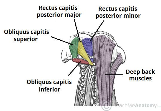
This triangle can be further divided into the submandibular triangle, submental triangle, muscular triangle and carotid triangle.
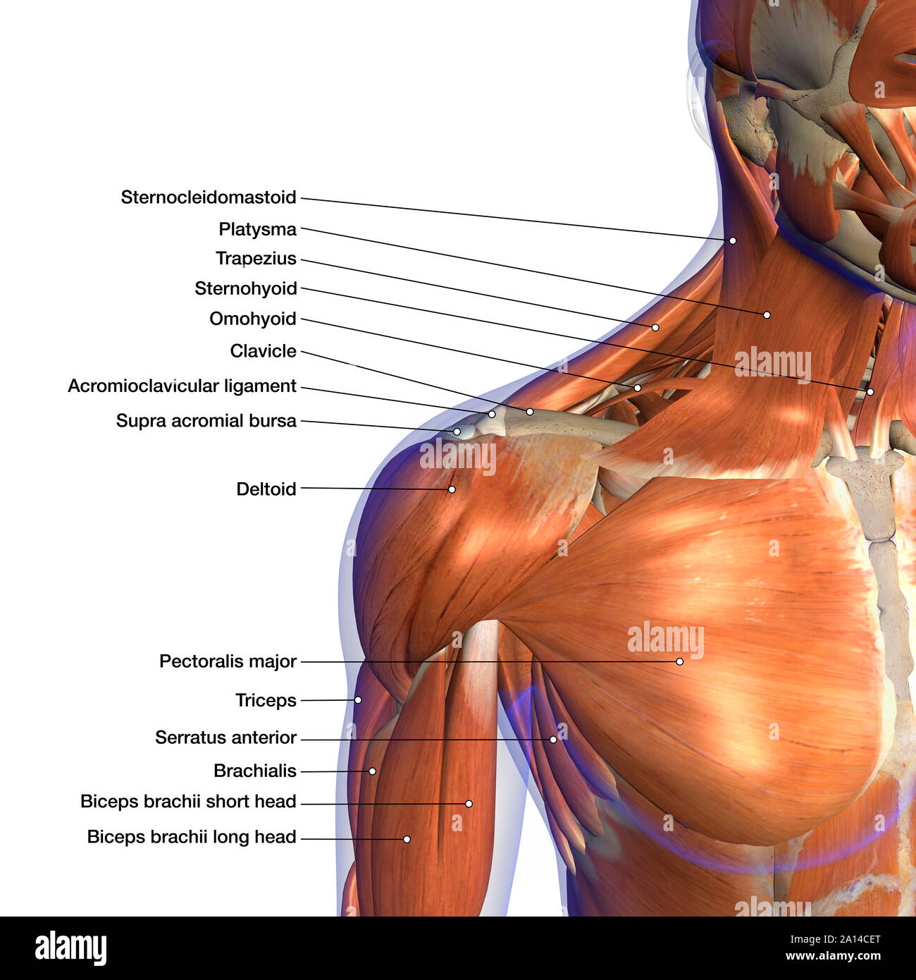
The neck muscles, including the sternocleidomastoid and the trapezius, are responsible for the gross motor movement in the muscular system of the head and neck. Just like the other joints in your body, your neck joints tend to wear down with age. Veins and arteries of the neck 9 photos of the veins and arteries of the neck activate javascript arteries in the neck diagram, common carotid artery branches, external carotid artery function, how many carotid arteries, left common carotid artery function, the left common carotid artery supplies blood to the. There are both right and left traps, and they are used to support your arms and shoulders. Learn vocabulary, terms, and more with flashcards, games, and other study tools. Head and neck muscles diagram in this image, you will find cranial aponeurosis, temporalis, occipitalis, masseter, sternocleidomastoid, trapezius, platysma, orbicularis oris, buccinator, zygomaticus, orbicularis oculi, frontalis in head and neck muscles diagram. Biology shoulder 3d illustration 3d rendering anatomical anatomy arm athletic biceps body bodybuilding brachialis bursa cgi chart deltoid diagram elbow fitness head health human human anatomy 3d. Article by steph dorworth | online fitness coach and physical therapist. Nerves in the neck, medically referred to as the cervical spine, help transmit information along the pathways of the central and peripheral nervous system, including sensory and motor skills processes.the cervical spine consists of eight different sets of nerves. Posted on september 10, 2014 by admin. The muscles on the front of the trunk help lift the arms and move. The anatomy of the neck and shoulders is very interesting. Stretches work, but you can also do simple exercises like the ones below.
Hold your phone higher up when you look at it, for example. Learn vocabulary, terms, and more with flashcards, games, and other study tools. Related posts of diagram of the neck anatomy veins and arteries of the neck. The trapezius, commonly referred to as the traps, are responsible for pulling your shoulders up, as in shrugging, and pulling your shoulders back during scapular retraction. A neck sprain or strain occurs when there is an injury to the soft tissues of the neck.
Labeled Anatomy Chart Of Neck And Shoulder Muscles On White Background Stock Photo Alamy from c8.alamy.com This triangle can be further divided into the submandibular triangle, submental triangle, muscular triangle and carotid triangle. Meanwhile, the splenius capitis is a much thicker, more broad muscle found on the back of the neck, which originates along the vertebrae in the neck and moves the base of the skull so that movements like shaking the head are possible. A neck sprain or strain occurs when there is an injury to the soft tissues of the neck. They can improve your neck strength and your range of motion. Head and neck muscles diagram in this image, you will find cranial aponeurosis, temporalis, occipitalis, masseter, sternocleidomastoid, trapezius, platysma, orbicularis oris, buccinator, zygomaticus, orbicularis oculi, frontalis in head and neck muscles diagram. Working in pairs on the left and right sides of the body, these muscles. The back's muscles start at the top of the back (named the cervical vertebrae) and go to the tailbone (also named the coccyx). You can work your neck muscles like any other muscles.
The muscles of the neck run from the base of the skull to the upper back and work together to bend the head and.
The pain can either pop up spontaneously (active) or when the trigger point is pressed (latent). Thank you for your support. There are anterior muscles diagrams and posterior muscles diagrams. The muscles on the front of the trunk help lift the arms and move. It is composed of three parts: Veins and arteries of the neck 9 photos of the veins and arteries of the neck activate javascript arteries in the neck diagram, common carotid artery branches, external carotid artery function, how many carotid arteries, left common carotid artery function, the left common carotid artery supplies blood to the. Our latest youtube film is ready to run. The back's muscles start at the top of the back (named the cervical vertebrae) and go to the tailbone (also named the coccyx). Learn vocabulary, terms, and more with flashcards, games, and other study tools. Learn vocabulary, terms, and more with flashcards, games, and other study tools. Our latest youtube film is ready to run. These critical parts of the upper body are very prone to developing pain because the position of all the bones in the neck and shoulders are completely dependent on the balance and alignment of the muscles and fascia that lash them together and allow for movement between them. There are both right and left traps, and they are used to support your arms and shoulders.